Sal

joined 3 years ago
MODERATOR OF
[–] Sal@mander.xyz 2 points 2 weeks ago (2 children)

Always happy to talk about molecules interacting with light! 😄

This is an aspect of lichen I hadn’t really put much thought into before now.

I have some background is in studying how light interacts with molecules, so I probably put more thought and emphasis on these things than average.

Its been in storage for a couple of months so I may try to re-hydrate it a bit before lighting it up during the night.

That's cool! When keeping a collection, can you keep them alive for a long time dry?

[–] Sal@mander.xyz 5 points 2 weeks ago* (last edited 2 weeks ago) (4 children)

Not sure how I managed to never hit this species with UV. I would describe the colour as a bright, hot, lipstick pink. I am unsure if this lichen is actually fluorescing or if something else to do with how the pigments show up under UV light - maybe @Sal@mander.xyz would know. Picture doesn’t quite do it justice.

You are pointing a UV lamp at it which probably sends out 365 nm or 395 nm photons. The lichen is shooting back photons with a broad range of wavelengths, and a lot of ~600 - 750 nm ones (red). So, the UV photons had to be "captured" by some molecular system, the system dissipated some energy, and then re-radiated some of these longer-wavelength photons.

The general term that covers the many different possibilities is "photoluminescence". In this case we can say for sure that the lichen exhibits "UV-induced photoluminescence", because it is re-emitting lower energy (longer-wavelength) photons. It is common to make the connection "photoluminescence" = "fluorescence", but technically fluorescence makes specific claims about how the light is re-emitted (singlet -> singlet emission), and it is not the only luminescence process. Other examples of luminescence are phosphorescence from a triplet state and luminescence via charge-recombination. So, to call it "fluorescence" in the strict sense we need know what the exact pathway is.

That said, when it comes to biological pigments fluorescence is generally the most common pathway. Triplets that live long enough to produce light are generally undesirable as they can react indiscriminately with molecules inside of the cell as well as produce reactive oxygen species, and good phosphorescent materials often combine metals and heavy atoms that are not as abundant in living tissue.

So, knowing nothing else, and seeing that red light comes out when you shine UV/blue light on a lichen, it is generally fair to call it "fluorescence".

Now, if we discuss this specific lichen... I have looked it up and it does get interesting! Do you have it with you? I suspect that its fluorescence might be different during the day than during the night.

I can find online two significant fluorescent components: parietin, which produces the fluorescent yellow pigment, and Chlorophyll a/b, which produces red fluorescence. There is an interesting paper exploring the idea that one functional purpose of parietin's fluorescence is that it can transfer energy to the algae to boost their photosynthesis. Their conclusions in the paper is that the idea is not supported by the evidence, so, a "negative result". It is a fun example of the type of research that is performed in photobiology and also an example to show that even negative results can be interesting enough to be published!

As for the difference between day and night - if what you see is a combination of the fluorescence of parietin and chlorophyll, then the color might change with the day/night cycle. Photosynthetic organisms regulate the flow of excess photon energy towards a safe non-radiative dissipation pathway in response to light. This is called the non-photochemical quenching pathway, and during the day this pathway tends to be active. During the night there is little light, and so this protective pathway shuts-off. This allows more of the absorbed photon energy to flow into the radiative fluorescence pathway, increasing the red fluorescence. You can actually see this easily with plants - you can dark adapt a leaf and then compare its fluorescence with that of a leaf that is being exposed to a bright light. The dark-adapted one will usually show significantly more red fluorescence.

This time you did ask, so I won't apologize for my essay 😆 But I am a bit sorry I didn't have the time to make it shorter.

[–] Sal@mander.xyz 7 points 2 weeks ago

No. I think they both lose more than they gain here. It doesn't make sense as a strategy. Ego clash is a simple explanation.

[–] Sal@mander.xyz 2 points 2 weeks ago (3 children)

Too bad you don't get to bring your equipment, but at least you will get to see them :D Good luck finding some wild ancestors!

[–] Sal@mander.xyz 2 points 2 weeks ago

Great find, congratulations!!

[–] Sal@mander.xyz 6 points 2 weeks ago (5 children)

Enjoy your holidays!! 🕊️

[–] Sal@mander.xyz 4 points 2 weeks ago (1 children)

Hi! I’ve looked through /r/shittyaskscience and I think it leans too far into jokes with very little actual science content. The idea behind mander is to support specific, niche science-related communities, so a general joke-focused community doesn't really fit.

For 'science_memes', the mod is a very capable superstar and I agree with their vision of memes as a laid-back way to connect people to science. It’s plausible that a community like 'shittyaskscience' could achieve something similar, but honestly I think science_memes already covers that space well.

As for !askscience - it simply hasn’t been created yet. It would be more fitting than 'shittyaskscience', but I still prefer encouraging people to ask lichen questions in the lichen community, mushroom questions in the mushroom community, chemistry questions in the chemistry community, and so on. I support content flowing toward niche communities rather than having a centralized place for general questions. A general community would be more popular, but popularity isn’t a goal, and it works against the underlying philosophy. Niche spaces may be smaller, but they offer much better signal-to-noise for building meaningful connections.

[–] Sal@mander.xyz 2 points 2 weeks ago (1 children)

Unfortunately the article is behind a paywall... But I am curious, does this mean there won't be any new Cortex-Mxs microprocessors?

The summary says that Arm wants to "enter the chip design space". They weren't doing this already?

And, is the prospect of a proprietary chip an exciting tease when pitted against an open standard one? I am excited about RISC-V microprocessors precisely because they rely on an open standard, so I am curious to see what their angle here is. I tried to find a non-paywalled source but I couldn't find one.

[–] Sal@mander.xyz 4 points 2 weeks ago (1 children)

I learned of your first attempt shortly after it went offline and I was bummed that I missed out on listening in. Thank you for trying again, I am excited to see how it goes!

[–] Sal@mander.xyz 1 points 2 weeks ago (1 children)

Solar Maximum happens every 11 years or so, right? I will keep paying attention!

[–] Sal@mander.xyz 2 points 2 weeks ago

Wow, that is spectacular!

14
Epithelial cells (mander.xyz)
submitted 1 month ago* (last edited 1 month ago) by Sal@mander.xyz to c/microscopy@mander.xyz
 

I followed the Gram Staining tutorial from this video to prepare a sample of my cheek cells: https://www.youtube.com/watch?v=lMoT-FmhS6A

For preparing the staining solutions I purchased crystal violet, ethanol, potassium iodide, iodine, and an already prepared safranin solution from laboratorium discounter.

The slight 3D effect is achieved by displacing the filter holder to block the light coming from one direction and achieve oblique illumination to cast a shadow (https://www.youtube.com/watch?v=9btIpf5mjyA).

The image is post-processed using Rawtherapee to increase the contrast.

Here is another photo without using the oblique illumination trick, also post-processed with rawtherapee:


In trying to isolate Trebouxia from an Evernia lichen. I found that some of the cultures are contaminated by a what I think are rotifers. I am not sure of what kind of rotifer (or other organism) is the one pictured, so if anyone has some idea please let me know.

I also recorded a video of what I think are belloid rotifers feeding on the same lichen culture:

https://peertube.uno/w/uoSCNagVVmbuMcgXdVfPGR

I don't have much hope that the algae will survive this attack, but I might turn those jars into rotifer cultures.


I left a slide with some algae and rotifers sitting on the microscope. After it dried up I was able to see several of these flower-like shapes. Not a pattern that I had seen before, and I a don't know what about the drying process lead to this particular shapes forming.


I encountered this golden young pigeon today


Abstract

Non-contact mechanical control of light has given rise to optical manipulation, facilitating diverse light-matter interactions and enabling pioneering applications like optical tweezers. However, the practical adoption of versatile optical tweezing systems remains constrained by the complexity and bulkiness of their optical setups, underscoring the urgent requirement for advancements in miniaturization and functional integration. In this paper, we present innovations in optical manipulation within the nanophotonic domain, including fiber-based and metamaterial tweezers, as well as their emerging applications in manipulating cells and artificial micro-nano robots. Furthermore, we explore interdisciplinary on-chip devices that integrate photonic crystals and optofluidics. By merging optical manipulation with the dynamism of nanophotonics and metamaterials, this work seeks to chart a transformative pathway for the future of optomechanics and beyond.

54
submitted 1 month ago* (last edited 1 month ago) by Sal@mander.xyz to c/Lichen@mander.xyz
 

About two years ago I wanted to learn more about lichen. Since I cultivate mushrooms as hobby, I figured that attempting to isolate the wild lichen symbionts and then re-synthesizing the lichen would be a good way to learn about them. The experiment was not successful, in part because it is a long process and at one stage I did not give it the attention it needed. But at least I can share a bit of the process and observation.

Near where I live I can easily find Oakmoss (Evernia prunastri) and the Yellow Wall Lichen (Xanthoria parietina).

I collected small samples of each and placed very small pieces of them into many agar dishes. The idea here is that, coming from a wild source, contaminants will be present for sure. The process of isolation is an iterative process in which you observe the growth on the plate, pick out a small region where your target organism is growing strongly, and then move it into another plate.

This is an agar plate into which pieces of Evernia were added:

Wild cuture of evernia, contaminated

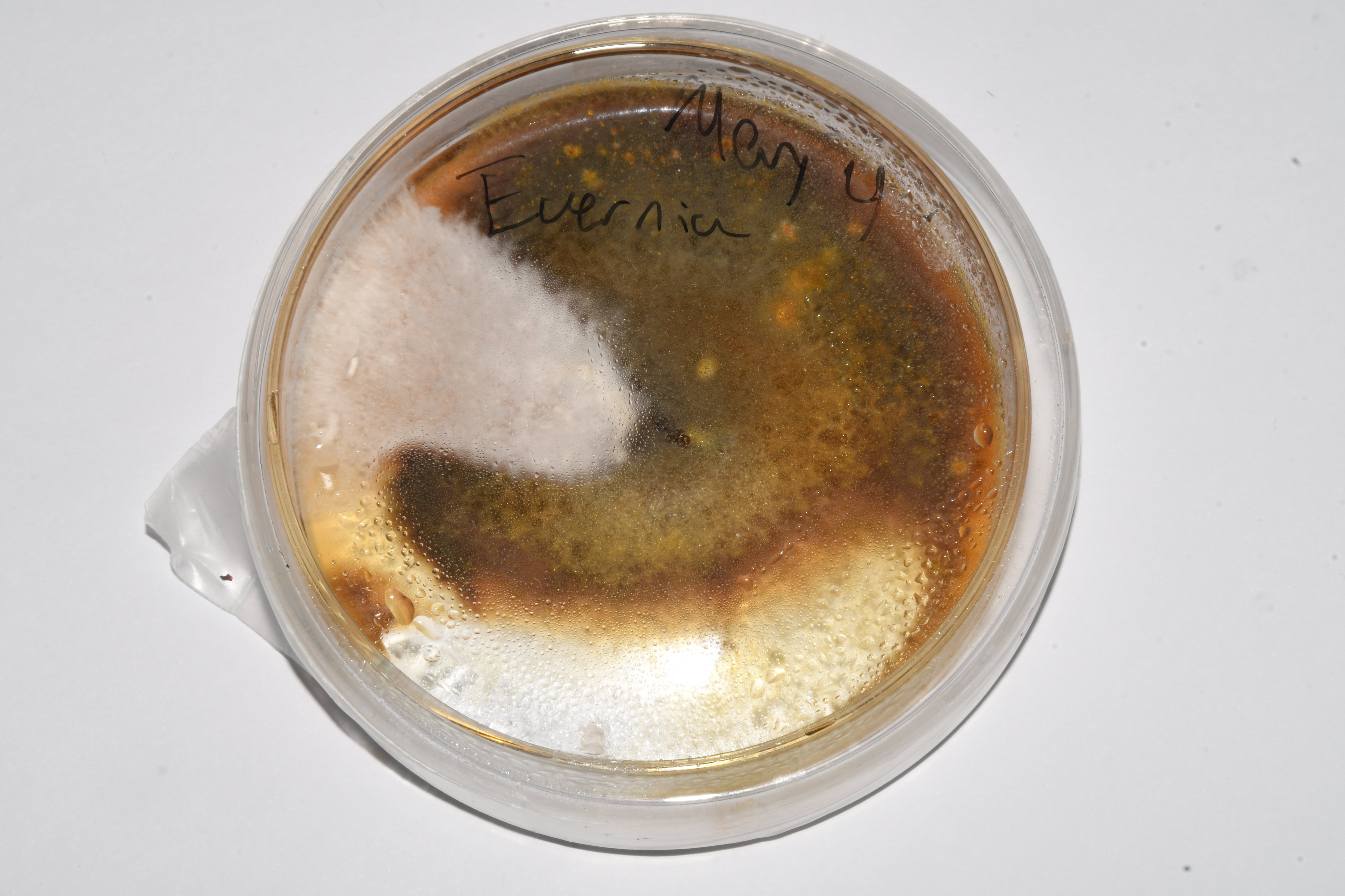
In this image you can see that there is a wedge with fluffy white mycelium, which was consistent with the morphology for the mycelium of this species as described in the literature. So, I would pick a tiny piece from this region and transfer into a new plate, to obtain a clean mycelium after 1 or 2 transfers:

Pure culture of evernia

This process was also performed for Xanthoria parietina.

After this step, I had plates with "clean" mycelium but not yet confirmation that the mycelium was truly the lichen's mycobiont. That is when microscopic identification comes into play. I was especially happy with the microscopic images I was able to get of the Xanthoria because they clearly show structures that are very characteristic structure of "septate, pluricellular, branched hyphae" described in the literature.

The microstructure of Xanthoria

At this point I now had the fungal component isolated in agar plates. This was for me the easy part because I am familiar with growing mushrooms. But to build a lichen we need two parts: a fungus and an algae.

From a search I could find that both lichen species may use algae of the genus Trebouxia as a symbiont. I placed small pieces of each lichen into water and made the following observations:

  • When placed in water, some of the algal cells become dislodged and float away

Photo: Algal cells floating away from a piece of Evernia prunastri

  • The algal cells can be seen held loosely in between the hyphae, rather than incorporated into the hyphal structure

Photo: Algal cells loosely bound to the hyphae of Evernia prunastri

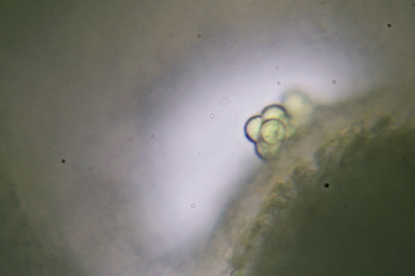
Photo: Cluster of algal cells bound to the surface of a hypha of Xanthoria parietina

  • Both species contained algar with similar if not identical round morphologies

Photo: Individual algal cell released from Xanthoria parietina

  • The round morphology is consistent with Trebouxia

.... So, at this point I had successfully isolated the fungal partner and had some evidence to suggest that the binding between the fungus and the algae is loose. Since I saw that the algae could be released from a lichen, an easy thing try was to attempt to transfer the algae from a living lichen to the mycelium culture.

This is a photo of that attempt. Small pieces of wild Evernia were placed on top of a mycelium culture that was already several weeks old.

This... Did not work at all. The culture eventually became contaminated and was thrown away.

The next attempt was to try to grow the algae separately. One method was to place the wild lichen into agar dishes with no added nutrients end expose the dishes to the sun. The logic here is that the algae will survive from photosynthesis while the rest of the species do not have the nutrients to thrive on. I tried this on about 15 plates, different light conditions, but nothing grew other than a few weakly growing contaminants in some of the dishes...

Another method was to place the lichen into a glass jars filled with water and place those by the window at different levels of shade.

In the meantime, I decided to grow "grain spawn" jars out of the mycobiont thinking that this would give me a lot of material to work with once the algae grew. Both fungi did colonize the grain well. However, those jars were abandoned and eventually, after several months of storage, became contaminated and I had to toss them away.

It has been almost two years and I just had a look now at how the algae in jars are doing.

In the Xanthoria jar I can see significant algal growth. As for the Evernia, the algae did not make it but it looks like the mycelium did, as it has created a floating white blob.

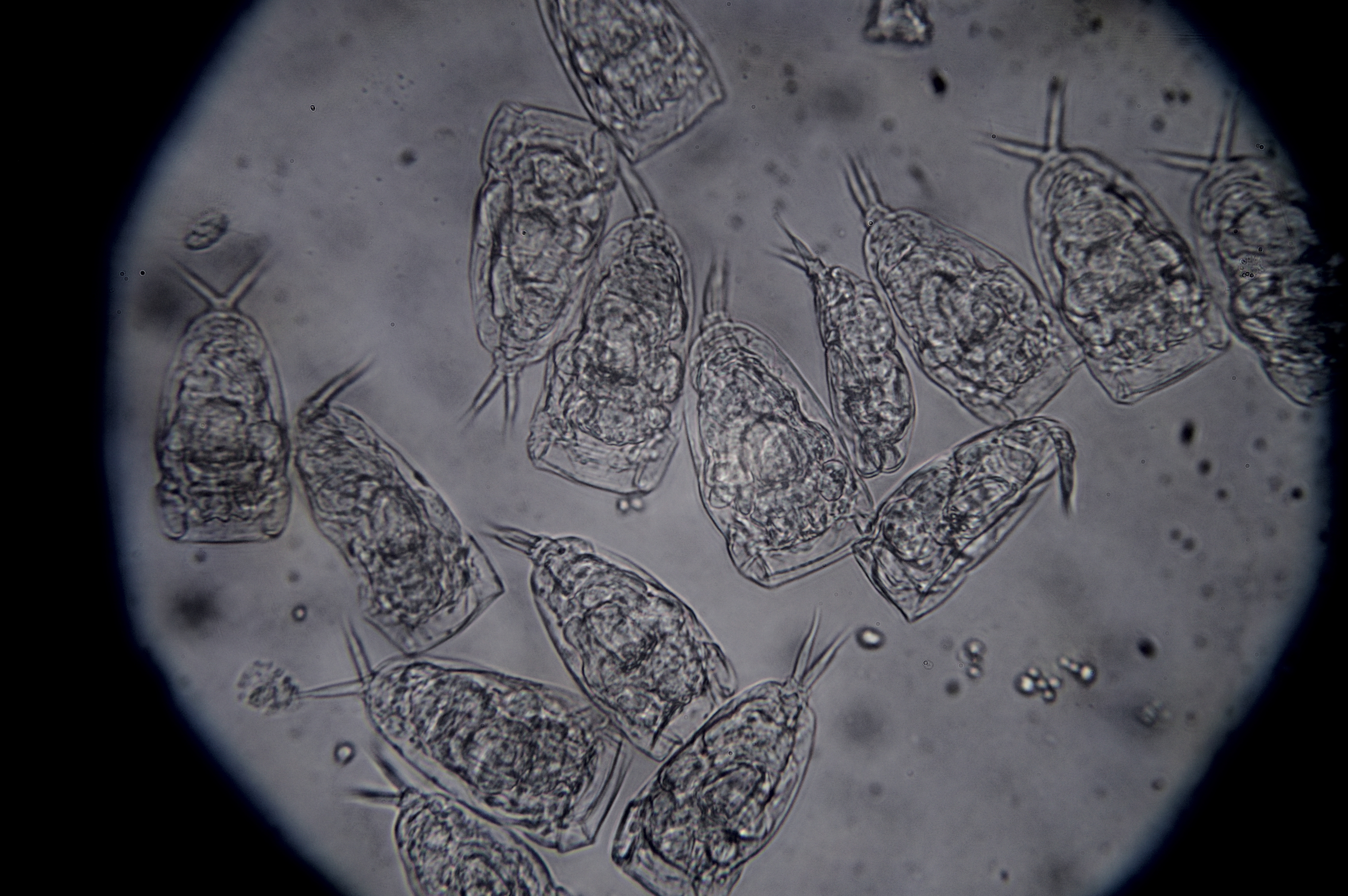
A few months after having the algae jar sitting by the window I analyzed the mixture under the microscope and did observe a large amount of Trebouxia.

Right now I have checked another sample, and, while I do see what looks like a bit of Trebouxia (marked with a red arrow), unfortunately most of the mixture now consists of other unidentified algal species. Since Trebouxia is not the dominant species, it would probably be easier to re-isolate the algae from the wild instead of trying to isolate it from this mixture.

I will give it a second try, and this time I will place more emphasis on culturing the algae first and keeping the cultures healthy and pure.

69
submitted 1 month ago* (last edited 1 month ago) by Sal@mander.xyz to c/electronics@discuss.tchncs.de
 

The SCD4x sensor from Sensirion measures CO₂, temperature, and humidity, and communicates these values via I²C.

The measurement principle for the CO2 is that of photoacoustic sensing. The fundamental principle is shown in the diagram below: shine light that the CO2 molecules absorb and use a microphone to listen to the pressure variations.

Principle of the CO2 sensor via photoacoustic sensing

I ordered a batch of SCD41 sensors from China for various projects, including fermentation, mushroom and plant cultivation, and field monitoring.

Since I had extras, I sacrificed one for macro photography. I removed the cover with a dremel and pliers, then cleaned the internals using isopropanol.

Here is my take:

The temperature and humidity are measured by Sensirion’s SHT40, seen as the black square at the bottom right. It’s likely accessed by the internal microcontroller over an internal I²C bus.

The pink square at the top left is a MEMS IR emitter. The SCD4x datasheet doesn’t specify the emission wavelength, but 4.3 µm is standard for NDIR-based CO₂ detection. A similar emitter example is this one from Microhybrid. These emitters usually produce broadband IR, with a 4.3 µm band-pass interference filter on top. The pink hue likely comes from this filter. Filters like these are critical to target CO₂ absorption while avoiding spectral overlap with other gases. For further reading, see Infratec's application note and Delta Optical Thin Film’s technical explanation.

The gold component labeled “o119 ANC” is the MEMS microphone, used to detect pressure waves caused by gas molecules absorbing pulsed IR light—this is photoacoustic sensing. The vibration excited by 4.3 µm light occurs at ~70 THz, far beyond acoustic detection. However, the IR source is pulsed at a modulation frequency (typically 20–60 Hz, e.g. 40 Hz), and the microphone detects the resulting pressure variations at this frequency. The principle is outlined in patent US 2024/0133801 A1.

An example of a compatible MEMS microphone is Infineon’s IM72D128V01, which supports frequencies down to 20 Hz.

The final main component is the metal-shielded package. It likely contains a microcontroller responsible for:

  • Driving the MEMS IR emitter with a modulated current (e.g., at 40 Hz)
  • Capturing and analyzing the MEMS microphone signal to extract the amplitude of acoustic pressure oscillations (proportional to CO₂ concentration)
  • Acting as an I²C master to retrieve temperature and humidity data from the SHT40
  • Acting as an I²C slave to provide CO₂, temperature, and humidity data to an external controller

Here are top and bottom views of the sensor cap:

Top view of cap of SCD41
Bottom view of cap of SCD41

The cap has a circular gas inlet. The white material covering it is likely a hydrophobic ePTFE membrane, which allows gas exchange while blocking liquid water.

I hope someone else finds this interesting too!


EDIT: After posted this, I searched online and I found a photo from someone who went a deeper than me and did expose the microcontroller: https://www.hackteria.org/wiki/CO2_Soil_Respiration_Chamber

This is the photo borrowed from that site:


58
submitted 1 month ago* (last edited 1 month ago) by Sal@mander.xyz to c/mander@mander.xyz
 

The developers of Lemmy are currently under-funded and are asking for donations.

Community funding of projects like Lemmy offers an alternative to the commercial system in which projects are funded by investors looking to make a profit in the long run.

Unfortunately, community funding is not easy. Sometimes it might be because many of the users are unable and/or unwilling to donate, but sometimes it might also be that it is not clear that donations are needed, or users may underestimate the impact of their "small" donation. I am glad that the developers are explicitly asking for help at this point, and hope that they find support to continue.

Cross-posting and pinning this here to help them spread their call for help. I recommend placing comments directly to the original post so that it is visible to them.

cross-posted from: https://lemmy.ml/post/29579005

An open source project the size of Lemmy needs constant work to manage the project, implement new features and fix bugs. Dessalines and I work full-time on these tasks and more. As there is no advertising or tracking, all of our work is funded through donations. Unfortunately the amount of donations has decreased to only 2000€ per month. This leaves only 1000€ per developer, which is not enough to pay my bills. With the current level of donations I will be forced to find another job, and drastically reduce my contributions to Lemmy. To avoid this outcome and keep Lemmy growing, I ask you to please make a recurring donation:

Liberapay | Ko-fi | Patreon | OpenCollective | Crypto

If you want more information before donating, consider the comparison with Reddit. It began as startup funded by rich investors. The site is managed by corporate executives who over time have become more and more disconnected from normal users. Their main goal is to make investors happy and to make a profit. This leads to user-hostile decisions like firing the employee responsible for AMAs, blocking third-party apps and more. As Reddit is a single website under a single authority, it means all users need to follow the same rules, including ridiculous ones like censoring the name "Luigi".

Lemmy represents a new type of social media which is the complete opposite of Reddit. It is split across many different websites, each with its own rules, and managed by normal people who actually care about the users. There is no company and no profit motive. Much of the work is carried out by volunteer admins, mods and posters, who contribute out of enthusiasm and not for money. For users this is great as there is no advertising nor tracking, and no chance of takeover by a billionaire. Additionally there are no builtin political or ideological restrictions. You can use the software for any purpose you like, add your own restrictions or scrutinize its inner workings. Lemmy truly belongs to everyone.

Dessalines and I work fulltime on Lemmy to keep up with all the feature requests, bug reports and development work. Even so there is barely enough time in the day, and no time for a second job. Previously I sometimes had to rely on my personal savings to keep developing Lemmy for you, but that can't go on forever. We partly rely on NLnet for funding, but they only pay for development of new features, and not for mandatory maintenance work. The only available option are user donations. To keep it viable donations need to reach a minimum of 5000€ per month, resulting in a modest salary of 2500€ per developer. If that goal is reached Dessalines and I can stop worrying about money, and fully focus on improving the software for the benefit of all users and instances. Please use the link below to see current donation stats and make your contribution! We especially rely on recurring donations to secure the long-term development and make Lemmy the best it can be.

Donate


There are a few people out there self-dosing with snake venom. The posted article is based on a study on the blood of one of these guys, Tim Friede, who has developed very effective antivenom in his blood after 20 years of self-dosing with a diverse array of snake venoms.

Vice did a few documentaries on Steve Ludwin, who is also self-immunizing. In one of these he answers questions about how he began and his motivation for doing what he does. In addition to producing anti-bodies he also believes that snake venoms have medicinal and anti-ageing properties - but these beliefs do not appear to be supported by any animal-based data as far as I can tell: https://www.youtube.com/watch?v=AcbqB0pFRPA

Self-dosing with snake venom is not something I would recommend. Generally a bad idea. But it is interesting to see the results and to learn about what motivates someone to do something like this.

34
submitted 1 month ago* (last edited 1 month ago) by Sal@mander.xyz to c/mander@mander.xyz
 

Sorry for the delay! The instance is now up-to-date running version 0.19.11

Important note: Moderators now have the ability to see what users upvote and downvote posts and comments within the communities that they moderate.

You can read the rest of the release notes here: https://mander.xyz/post/27810123


Abstract

For nearly 450 million years, mycorrhizal fungi have constructed networks to collect and trade nutrient resources with plant roots1,2. Owing to their dependence on host-derived carbon, these fungi face conflicting trade-offs in building networks that balance construction costs against geographical coverage and long-distance resource transport to and from roots3. How they navigate these design challenges is unclear4. Here, to monitor the construction of living trade networks, we built a custom-designed robot for high-throughput time-lapse imaging that could track over 500,000 fungal nodes simultaneously. We then measured around 100,000 cytoplasmic flow trajectories inside the networks. We found that mycorrhizal fungi build networks as self-regulating travelling waves—pulses of growing tips pull an expanding wave of nutrient-absorbing mycelium, the density of which is self-regulated by fusion. This design offers a solution to conflicting trade demands because relatively small carbon investments fuel fungal range expansions beyond nutrient-depletion zones, fostering exploration for plant partners and nutrients. Over time, networks maintained highly constant transport efficiencies back to roots, while simultaneously adding loops that shorten paths to potential new trade partners. Fungi further enhance transport flux by both widening hyphal tubes and driving faster flows along ‘trunk routes’ of the network5. Our findings provide evidence that symbiotic fungi control network-level structure and flows to meet trade demands, and illuminate the design principles of a symbiotic supply-chain network shaped by millions of years of natural selection.


Abstract

A newly designed optical aluminosilicate glass that supports femotsecond laser written ultra-low loss optical waveguides is presented in this paper. Propagation losses as low as −0.020 ± 0.003 and −0.037 ± 0.003 dB cm−1 at 1310 and 1550 nm, respectively, are enabled by engineering the glass composition. Raman, Brillouin and electron microscopies are used to understand the origins of femtosecond laser-induced refractive index changes.

view more: ‹ prev next ›